
Scientists in Virginia used light-sheet microscopy to video the goings-on of the larva’s nervous system
ASHBURN, Va. – Scientists at the Howard Hughes Medical Institute have imaged motor neuron activity in a millimetre-long larva.
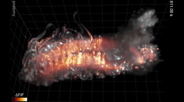
Their paper was published today in the open-access journal Nature Communications. Photographs were taken of the neurons of the larva, Drosophila melanogaster, five times per second, using penetrative lasers as part of a cutting edge technique called light-sheet microscopy.
The footage has been made available on YouTube l, and provides a fascinating insight into one of nature’s great miracles. Only the larva’s nervous system is shown, and areas of activity glow orange-red. The nerves are being artificially stimulated, as if the larva were crawling around.
[youtube id=”eTiSMC_fbSg” align=”center” mode=”normal” autoplay=”no” maxwidth=”600px”]
Philipp Keller, a participating scientist, said: “By imaging different parts of the nervous system at the same time, we can see how behaviours are controlled and then build models of how it all works.”
The study builds on research which involved even smaller organisms like nematode worms. By imaging not just the brain, but also the nerve cord, scientists can better see how the two work together. The team has now moved on to adult flies, zebrafish, and mouse embryos.
By Robbie Carney