Neuroscientists and engineers at North Carolina’s Duke University have pioneered a method with which the effects of transcranial magnetic stimulation (TMS) on the brain can be measured. The Duke team has made it possible to measure the response of a single neuron to an electromagnetic charge–something that has not before been possible. The work offers the potential to improve and initiate novel TMS therapy approaches.

“This report focused on the innovative methodology that allowed us to record from single neurons,” Duke professor of biomedical engineering, electrical and computer engineering, and neurobiology and lead researcher on the team, Warren Grill, told The Speaker. The team was able to record an increase in a neuron’s firing rate in the wake of the short, rapidly varying magnetic field created by TMS. The increase in firing lasted approximately 100 ms after the TMS pulse, according to Grill.
The report, “Simultaneous transcranial magnetic stimulation and single-neuron recording in alert non-human primates,” was authored by Jerel K Mueller, Erinn M Grigsby, Vincent Prevosto, Frank W Petraglia III, Hrishikesh Rao, Zhi-De Deng, Angel V Peterchev, Marc A Sommer, Tobias Egner, Michael L Platt, in addition to Grill, was published in Nature and was supported by a Duke Institute for Brain Sciences Research Incubator Award and by a grant from the National Institute of Neurological Disorders and Stroke of the National Institutes of Health.
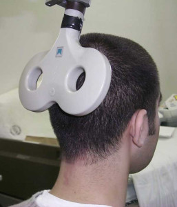
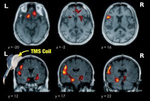
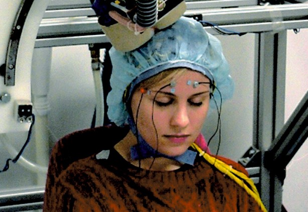
 Transcranial magnetic stimulation is a widely-used procedure wherein electromagnetic coils are held up to the skull and short electromagnetic pulses are run through the coil. It has long been understood that neurons react to TMS, and the procedure has been used to treat psychiatric disorders, substance abuse and other health conditions. Although preferable to other treatment methods because TMS is noninvasive, its mechanisms have always been poorly understood, making improvements difficult.
In part, the barrier to understanding the mechanisms of TMS is due to the difficulty of measuring neural responses during the procedure. The neural response is electric,and the current charging the TMS bears an overwhelmingly stronger electric charge.
Grill said of the difficulty in understanding TMS without measuring its effects, “Nobody really knows what TMS is doing inside the brain, and given that lack of information, it has been very hard to interpret the outcomes of studies or to make therapies more effective. We set out to try to understand what’s happening inside that black box by recording activity from single neurons during the delivery of TMS in a non-human primate. Conceptually, it was a very simple goal. But technically, it turned out to be very challenging.”
Although thousands of times smaller than the charge of the TMS, the neural response can be measured by the research team’s hardware. The team also overcame the obstruction posed by the recording device, which also emitted an electric current.
 “Studies with TMS have all been empirical,” said Grill. “You could look at the effects and change the coil, frequency, duration or many other variables. Now we can begin to understand the physiological effects of TMS and carefully craft protocols rather than relying on trial and error. I think that is where the real power of this research is going to come from.”
The Duke team’s research is open to anyone with a lab, according to the researchers. “[A]ny modern lab working with non-human primates and electrophysiology can use this same approach in their studies,” said Grill. The team said they hope others would pursue this line of research, and contribute to improvements in TMS therapy.
“This research will allow us first to quantify and understand the effects of TMS on neurons, and subsequently to design novel approaches, including stimulation waveforms and stimulation coil design to amplify or modify those effects,” Grill told us.
By Day Blakely Donaldson
